
Understanding
Hip Dysplasia
Healthy Hips. Happy Dogs. Happy Families.
Hip dysplasia isn’t simply a hip that “looks bad” on an X-ray. It begins with abnormal development and instability within the joint. Understanding what happens inside a healthy hip joint opposed to a loose hip joint makes it easier to understand why breeders test hips—and what those results can tell us.
Our goal with this page is to make hip health easier to understand for both families and breeders. Hip dysplasia is often discussed in terms of scores, grades, and X-rays, but those results make much more sense when we understand what is actually happening inside the joint. By looking at the mechanics of a healthy hip, joint laxity, subluxation, abnormal loading, and the changes that can develop over time, we hope to make hip testing less abstract and more meaningful.
We’ll also share real radiographs from dogs in our own program to show what these differences can look like in real life, and how tools such as OFA and PennHIP help us gather information for thoughtful breeding decisions. Throughout the page, we’ll link directly to scientific research and official veterinary and testing resources so readers can explore the evidence and source material for themselves.
HIP DYSPLASIA EDUCATION
Terms to Know
A Quick Reference To Common Terms Used On This Page
Understanding these key terms can make it easier to follow the information, compare results and have meaningful conversations about hip health.
Joint Loading
The forces placed on and transmitted through a joint during weight bearing and movement. In a stable hip, these forces are distributed across a broad area of the joint surface.
Joint Laxity
Excessive looseness or movement within a joint. In the hip, laxity allows the femoral head to move farther within or away from the acetabulum than it should.
Congruity
How well the shape and position of the femoral head and acetabulum match one another when the joint is seated.
Subluxation
Partial displacement of the femoral head from its normal position within the acetabulum. Unlike a complete dislocation, the femoral head remains partially within the socket.
Acetabular Coverage
How much of the femoral head is contained or covered by the acetabulum. Coverage and laxity are related aspects of hip function, but they are not the same thing.
Articular Cartilage
The smooth tissue covering the surfaces of the bones within the joint. It reduces friction and helps distribute forces during movement.
Osteophytes
New bone growth, often called bone spurs, that can develop around the margins of a joint in response to chronic instability and osteoarthritis.
Osteoarthritis (OA)
Progressive degeneration and remodeling of a joint involving cartilage, bone and other joint tissues. In hip dysplasia, OA can develop as a consequence of chronic instability and abnormal joint mechanics. Also known as Degenerative Joint Disease (DJD).
1 - What should a hip look like?
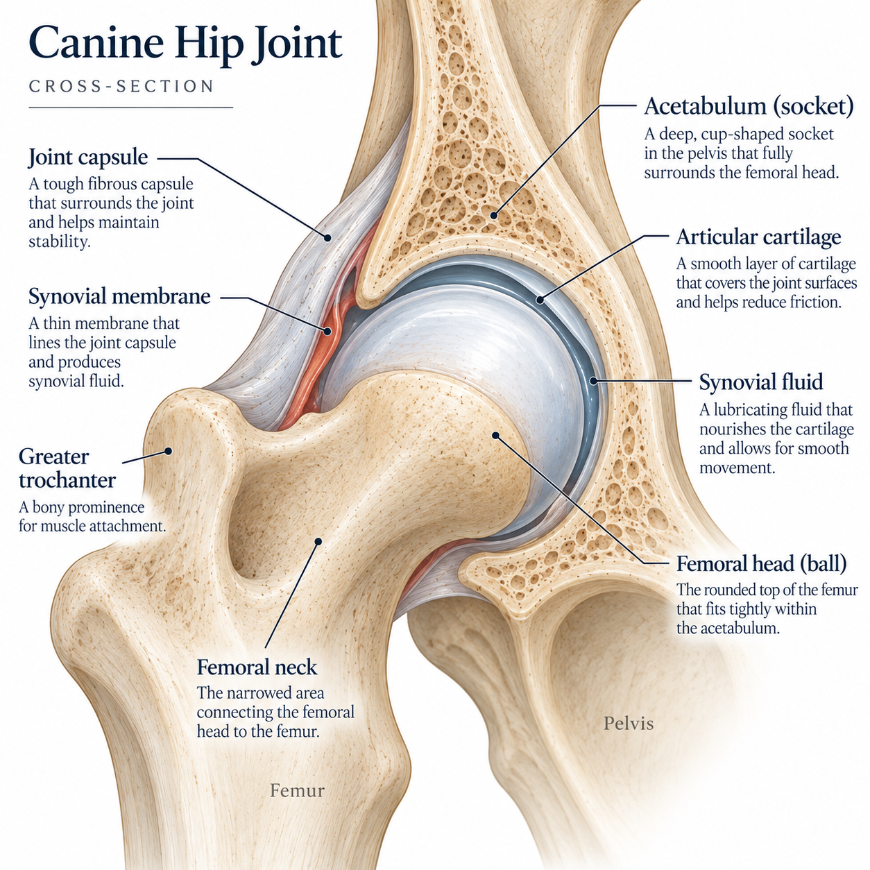
The hip is a ball-and-socket joint designed to provide both stability and a wide range of movement. The rounded femoral head, or ball, sits within the acetabulum, the cup-shaped socket formed by the pelvis. In a well-formed hip, the socket provides good coverage of the femoral head, helping keep it securely seated while spreading the forces of movement and weight bearing across a broad surface.
The surfaces of both the femoral head and acetabulum are covered with smooth articular cartilage. This cartilage creates a low-friction surface that allows the bones to glide smoothly against one another while also helping absorb and distribute forces through the joint.
Surrounding the hip is the joint capsule, a strong sleeve of connective tissue that encloses the joint and contributes to its stability. The inside of the capsule is lined by the synovial membrane, which produces synovial fluid. This fluid lubricates the joint and helps nourish the cartilage.
The femoral neck connects the femoral head to the rest of the femur and helps position the ball correctly within the socket. Together, the shape of the bones, depth of the socket, cartilage, joint capsule and surrounding soft tissues allow the hip to remain stable while still moving freely.
When all of these structures work together, pressure is distributed over a large area of the joint rather than concentrated in a few places. That relationship between stability, coverage and load distribution is important for understanding what happens when a hip is excessively lax.
A well-functioning hip is stable, congruent, and allows for smooth pain-free movement.
2 - What is Hip Laxity and subluxation?
Hip laxity refers to looseness or excessive movement within the hip joint. Instead of the femoral head remaining snugly seated within the acetabulum, a lax hip allows the ball to move farther away from the socket than it should.
Some movement within a joint is normal and necessary. The problem occurs when there is enough looseness that the femoral head can subluxate, meaning it shifts partially out of its normal position within the socket, particularly as forces are placed on the joint.
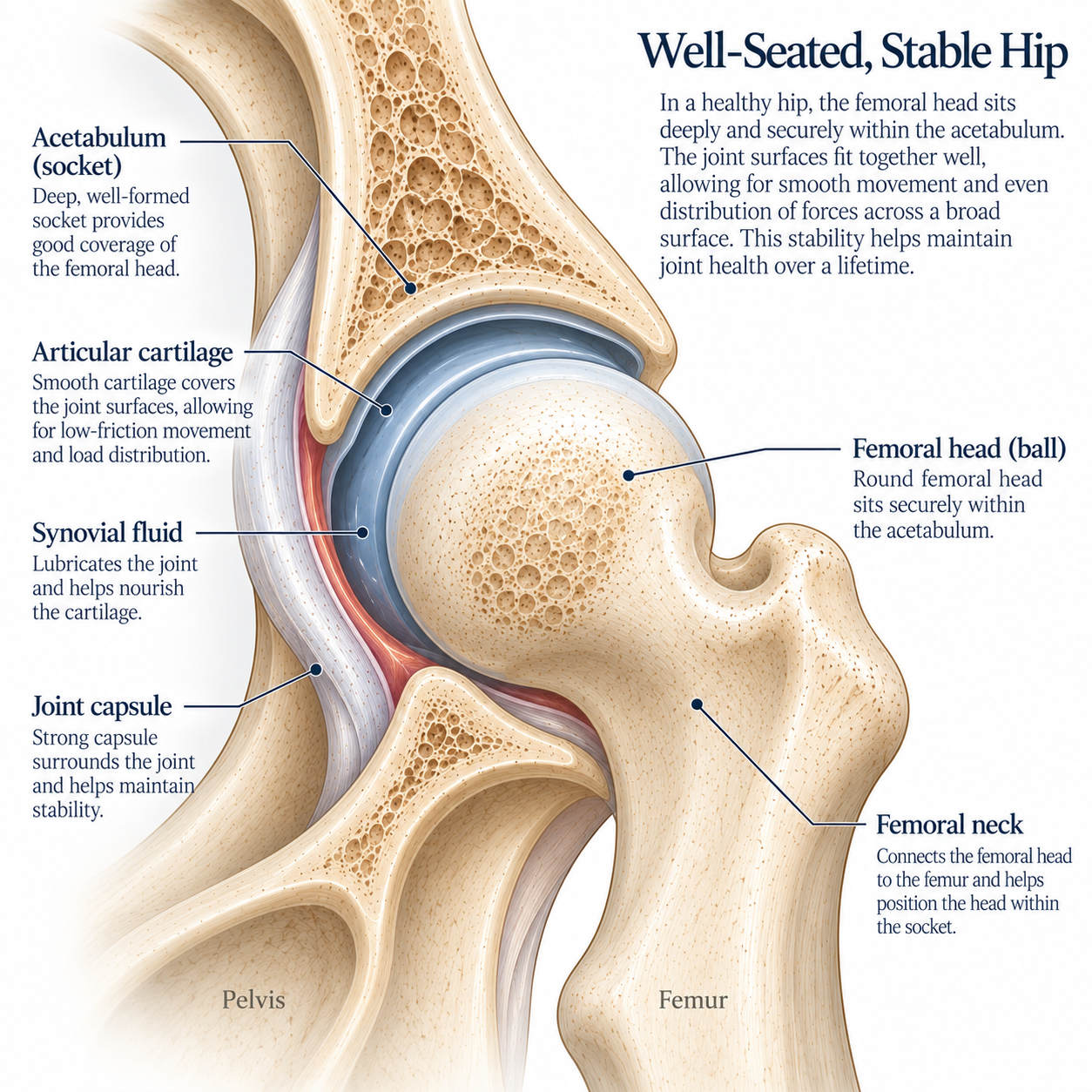
The more the femoral head is able to shift within the socket, the more joint stability and normal load distribution can be affected. A loose hip doesn’t just move more. It bears weight differently.
Why does laxity matter?
A healthy hip works partly because the femoral head and acetabulum have a close, congruent fit. This allows the forces created by running, jumping and everyday movement to be distributed across a broad area of cartilage.
When the hip is lax, that relationship changes. As the femoral head shifts within the socket, less of its surface may be supported by the acetabulum. The same forces are then concentrated over a smaller area of the joint rather than being evenly distributed.
That abnormal movement and loading can place repeated stress on the articular cartilage, joint capsule and underlying bone. The joint capsule may stretch and thicken, cartilage can become damaged, and inflammation can develop within the joint.
Over time, the body responds to chronic instability by trying to remodel and stabilize the joint. New bone may form around the margins of the joint as osteophytes, while the shape of the femoral head, femoral neck and acetabulum can gradually change. Typically, the femoral head and acetabulum will flatten and create rough edges, with thickening of the femoral neck.
This is how an initially loose, otherwise relatively normal-looking young hip can eventually develop the structural changes we recognize as osteoarthritis associated with canine hip dysplasia.
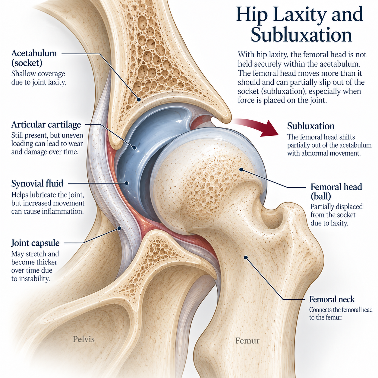
The changes we recognize as hip dysplasia can develop over time as the joint responds to chronic instability and abnormal loading.
Examples of Tight, Well-Seated Hips
The radiographs below are from dogs in our own breeding program that have received passing OFA evaluations or favorable PennHIP results. They provide real-world examples of hips with good stability and well-seated femoral heads. Because no two hips are identical, you’ll notice some natural variation in shape, depth, and coverage even among dogs with good hip evaluations.
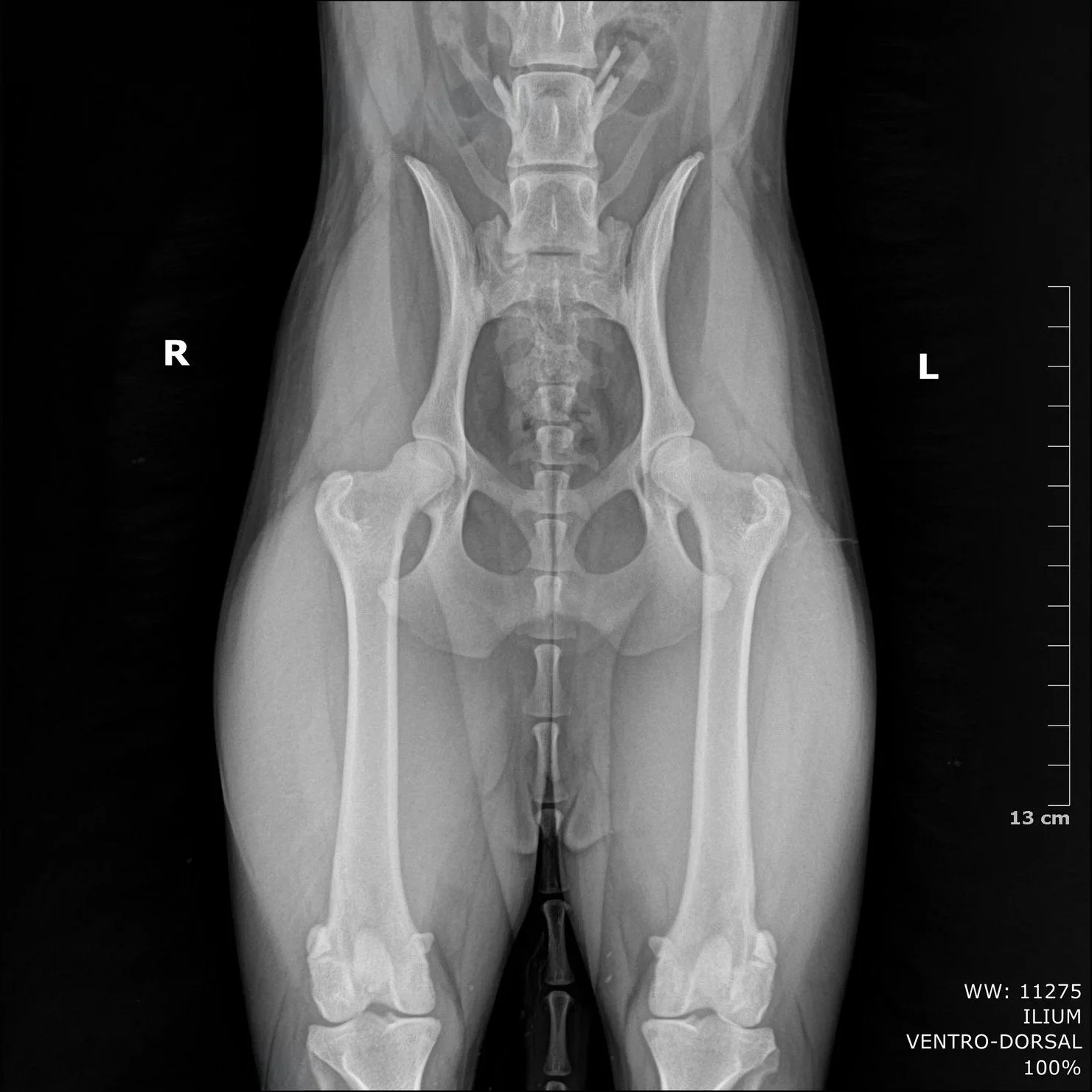
OFA Excellent
Polly - OFA extended view
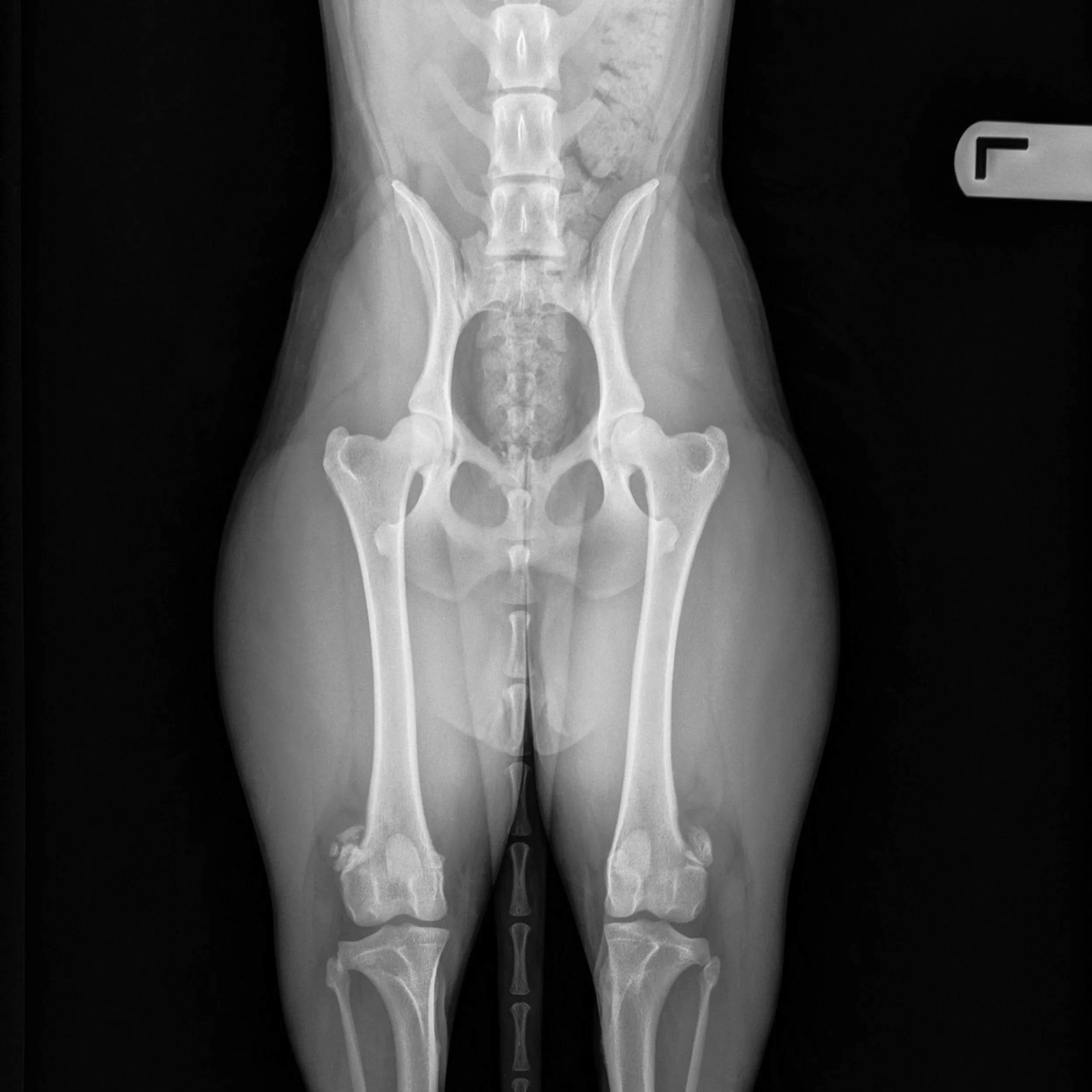
OFA Excellent
Talulah - OFA extended view

PennHIP .15/.15
Emmet - PennHip Extracted View
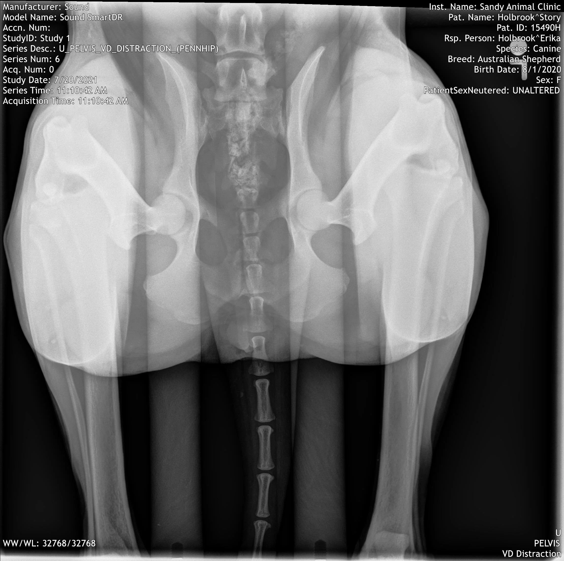
PennHIP .26/.25
Story - PennHip Extracted View
Examples of Hip Instability and Arthritic Change
The radiographs below include dogs from our own program as well as dogs from other breeding programs whose images have been shared with us for educational purposes. They show real-world examples of hips with varying degrees of poor seating, subluxation, remodeling, and arthritic change. Not every hip shown has the same findings or severity, but together they help illustrate how instability and degenerative changes can appear on radiographs.
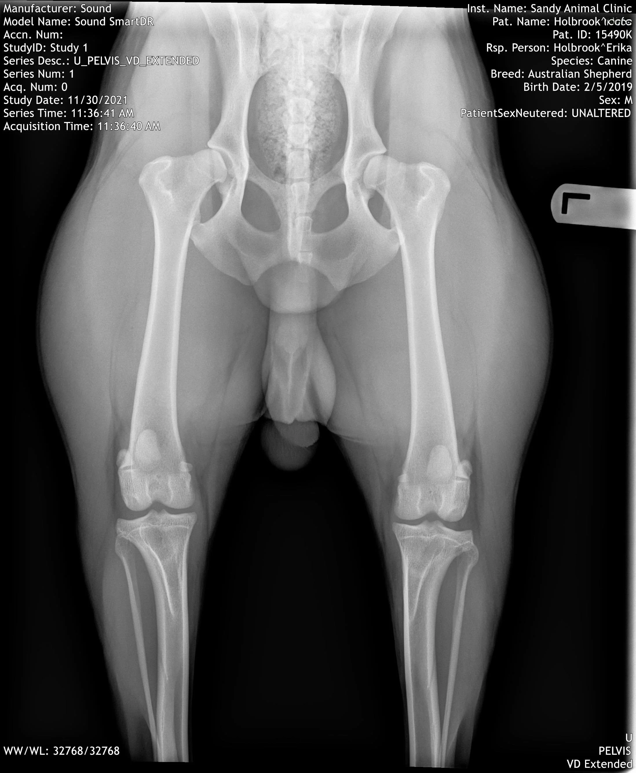
PennHIP .40/.43
PennHip extended view - Moderate OA
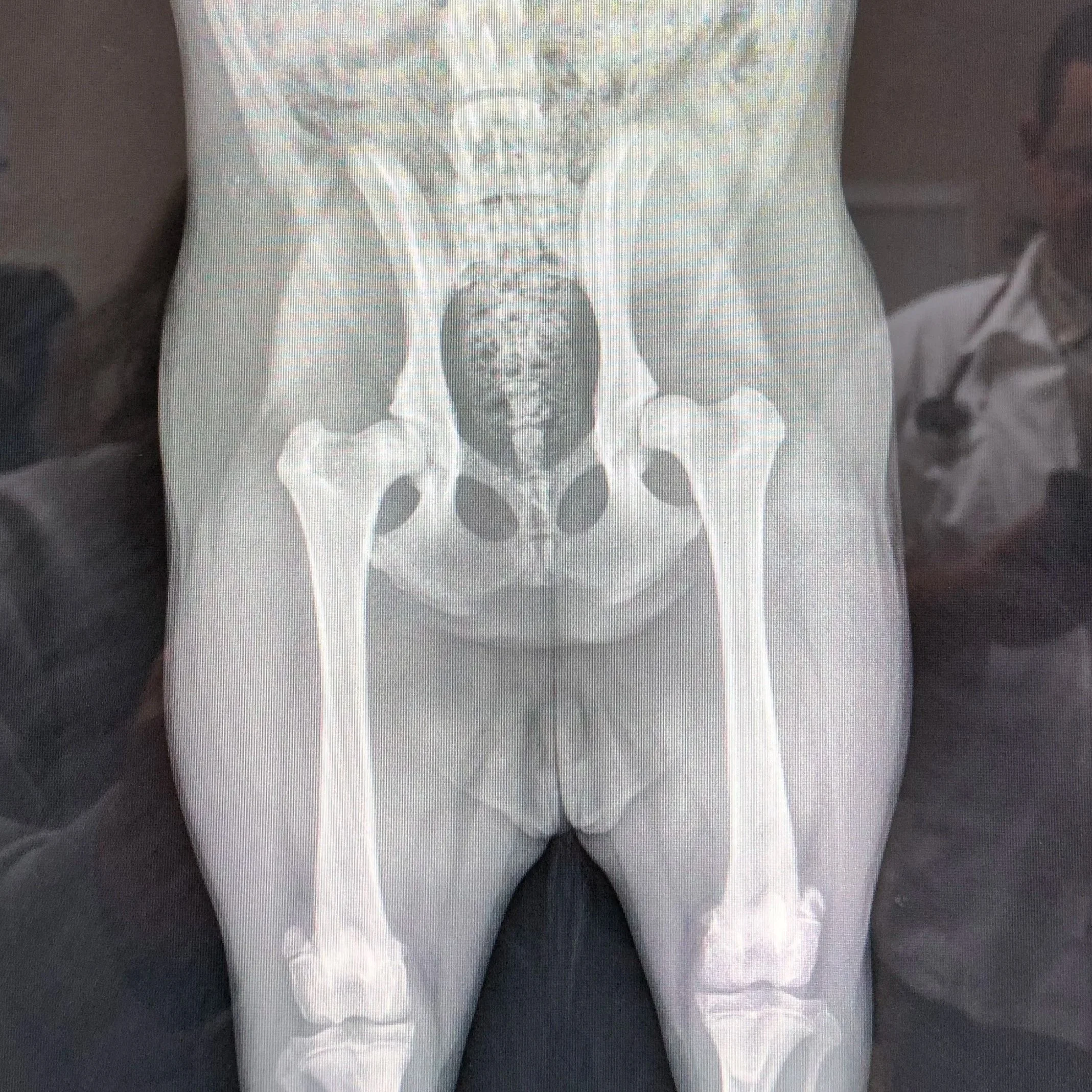
Severe OA
Crystal - Extended View Diagnostic RAD
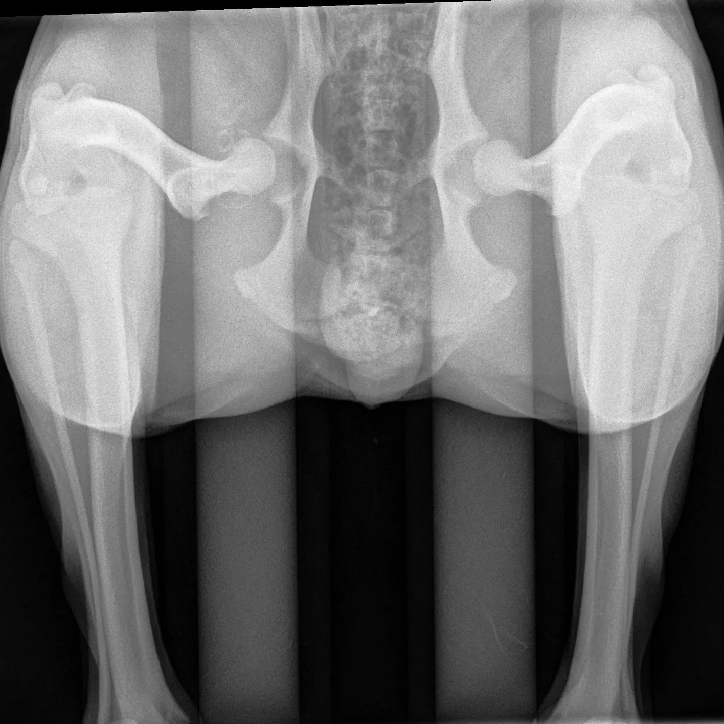
PennHIP .84/.79
Jupiter - PennHip Extracted View Mild OA
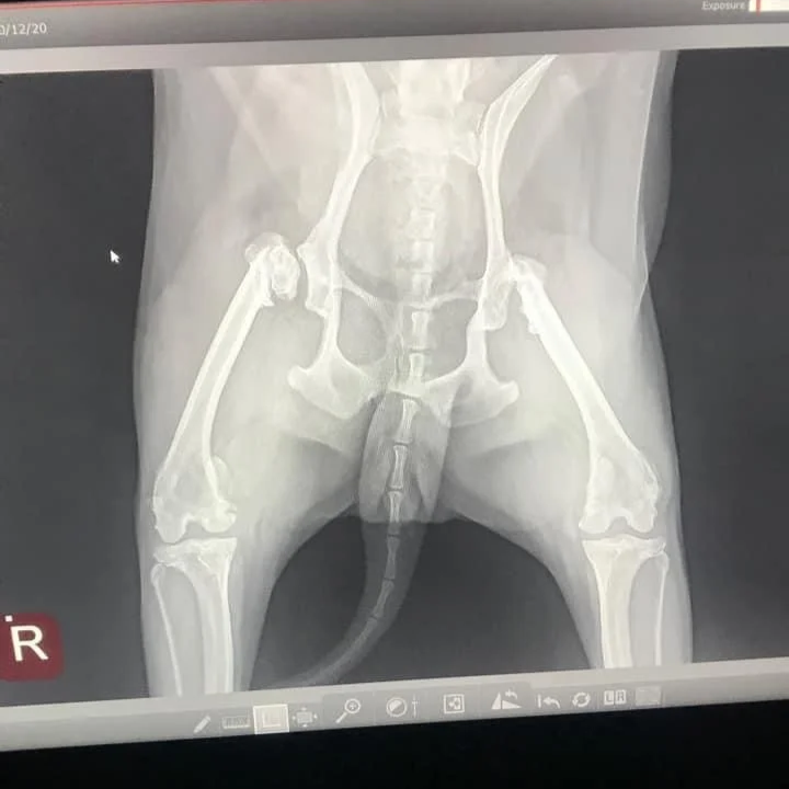
Severe OA
Shared Image - OFA/PennHip Extended View
3 - How the Body responds to abnormal joint mechanics
In the previous section, we looked specifically at hip laxity and subluxation and how excessive movement of the femoral head can alter the way forces are carried through the joint. But hip health can be evaluated in different ways, and not every screening method places the same emphasis on the same characteristics.
OFA and PennHIP approach hip evaluation differently. OFA evaluates the overall radiographic appearance and conformation of the hip on an extended view, including factors such as congruity, seating, acetabular coverage, subluxation and evidence of degenerative change. PennHIP uses three radiographic views and places particular emphasis on objectively measuring passive hip laxity through the Distraction Index (DI), which is used to estimate a dog's risk of developing hip osteoarthritis. OFA PennHip
These differences are important because hip shape, coverage, congruity and laxity are related, but they are not interchangeable measurements. A hip that receives a lower OFA passing grade based partly on its appearance or coverage does not necessarily have a high PennHIP DI, just as an extended radiograph that appears well seated may not reveal the amount of passive laxity demonstrated on a PennHIP distraction view.
What ultimately matters to the joint itself is how it functions mechanically over time. When abnormal movement or loading repeatedly stresses the tissues of the hip, the joint can begin to respond and remodel.
Hip shape, coverage, congruity and laxity describe different characteristics of the joint. What matters over time is how those characteristics affect the way the hip moves and carries load.
The Body Tries To Stabilize The Joint

Bone is living tissue that continually adapts to the forces placed upon it. When a hip experiences chronic instability or abnormal loading, the body responds by attempting to reinforce and stabilize the joint. Unfortunately, this protective response can also permanently change the structure of the hip.
As abnormal forces continue, the bone beneath the articular cartilage can become thicker and denser, a process called subchondral sclerosis. At the margins of the joint, the body may begin laying down new bone. These new bony projections are called osteophytes, or bone spurs, and are one of the radiographic signs of osteoarthritis.
The shape of the joint can also gradually change. The femoral neck may thicken, the normally smooth margins of the femoral head and acetabulum may become irregular, and the femoral head can lose some of its round shape. With continued degeneration, both the ball and socket may become increasingly remodeled.
These changes aren't the body simply “building a bad hip.” They are, in part, the body's response to a joint experiencing abnormal mechanical stress. The goal is stability, but the result can be a joint that becomes thicker, less smoothly shaped and less capable of normal, low-friction movement.
Over time, this combination of cartilage degeneration, inflammation and bone remodeling is what we recognize as osteoarthritis (OA) or degenerative joint disease (DJD).
The arthritic changes visible on an X-ray are often the end result of a process that began much earlier inside the joint.
4 - How Does Hip Dysplasia Affect The Dog?
One of the challenging things about hip dysplasia is that the severity of the changes within the joint does not always match the severity of the dog's symptoms. Some dogs with significant laxity, remodeling, or osteoarthritis may remain active and appear comfortable for years. Others may show pain or changes in movement much earlier.
Dogs can also compensate remarkably well. Muscle strength, body condition, activity level, age, the degree of inflammation, and the individual dog's response to pain can all influence how noticeable the condition becomes. For this reason, a dog that appears completely sound and comfortable can still have abnormal hips, while another dog with less dramatic radiographic changes may be more symptomatic.
This is why we cannot determine hip health simply by looking at a dog, watching it move, or evaluating it from the outside. A dog may run, jump, compete, work, and show no obvious signs of discomfort while still having significant abnormalities within the hip joint. Radiographs allow us to see the underlying bones and evaluate joint structure, congruity, laxity, and/or degenerative changes that may otherwise remain hidden.
For breeders, this is especially important. Canine hip dysplasia has a heritable component, which means an apparently healthy dog's hip status can still matter to the next generation. Screening breeding dogs before they are bred gives us information we cannot obtain from appearance or movement alone and allows us to make more informed breeding decisions.
Unless a dog is showing symptoms, we cannot see hip health from the outside. Radiographic screening gives breeders information that appearance, movement, and performance alone cannot.
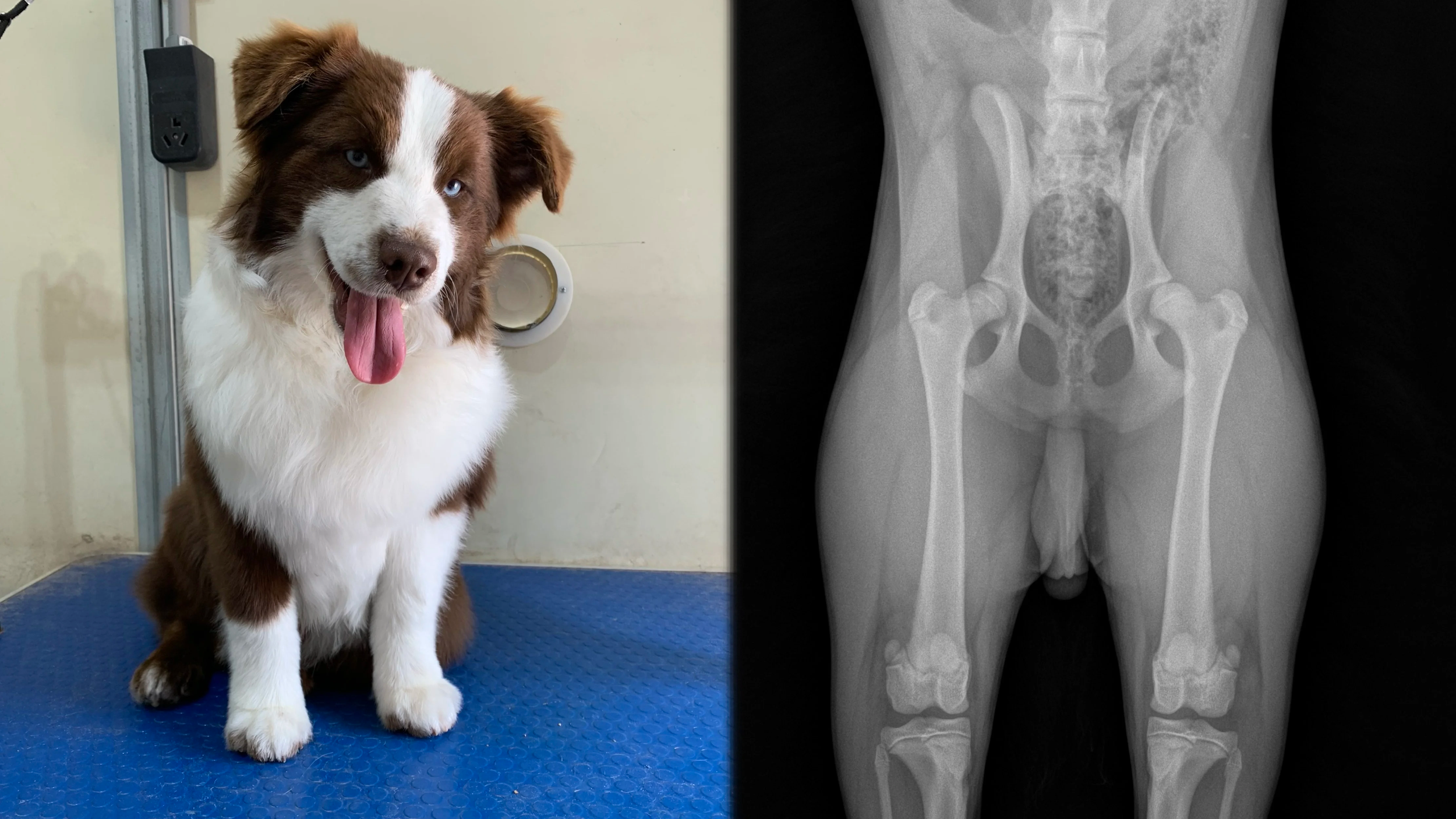
Jupiter - Diagnosed with severe hip laxity by PennHIP at 6 months old. Jupiter had never shown a single symptom, and we fully expected him to have good results. Instead, his PennHIP evaluation revealed some of the highest Distraction Index scores our veterinarian had seen.
Possible Signs Of Hip Dysplasia
Symptoms can be subtle, intermittent, or progressive. They may include:
Stiffness, particularly after rest or exercise
Difficulty getting up or lying down
Reluctance to jump, climb stairs, or get into a vehicle
Reduced endurance or willingness to exercise
Changes in gait or stride
“Bunny hopping” with the rear legs when running
Swaying or altered movement through the hindquarters
Shifting weight toward the front legs
Difficulty extending or flexing the hip
Loss of muscle mass in the hindquarters
Pain or sensitivity around the hips
Lameness that may be intermittent or persistent
Changes in activity or behavior that can reflect discomfort
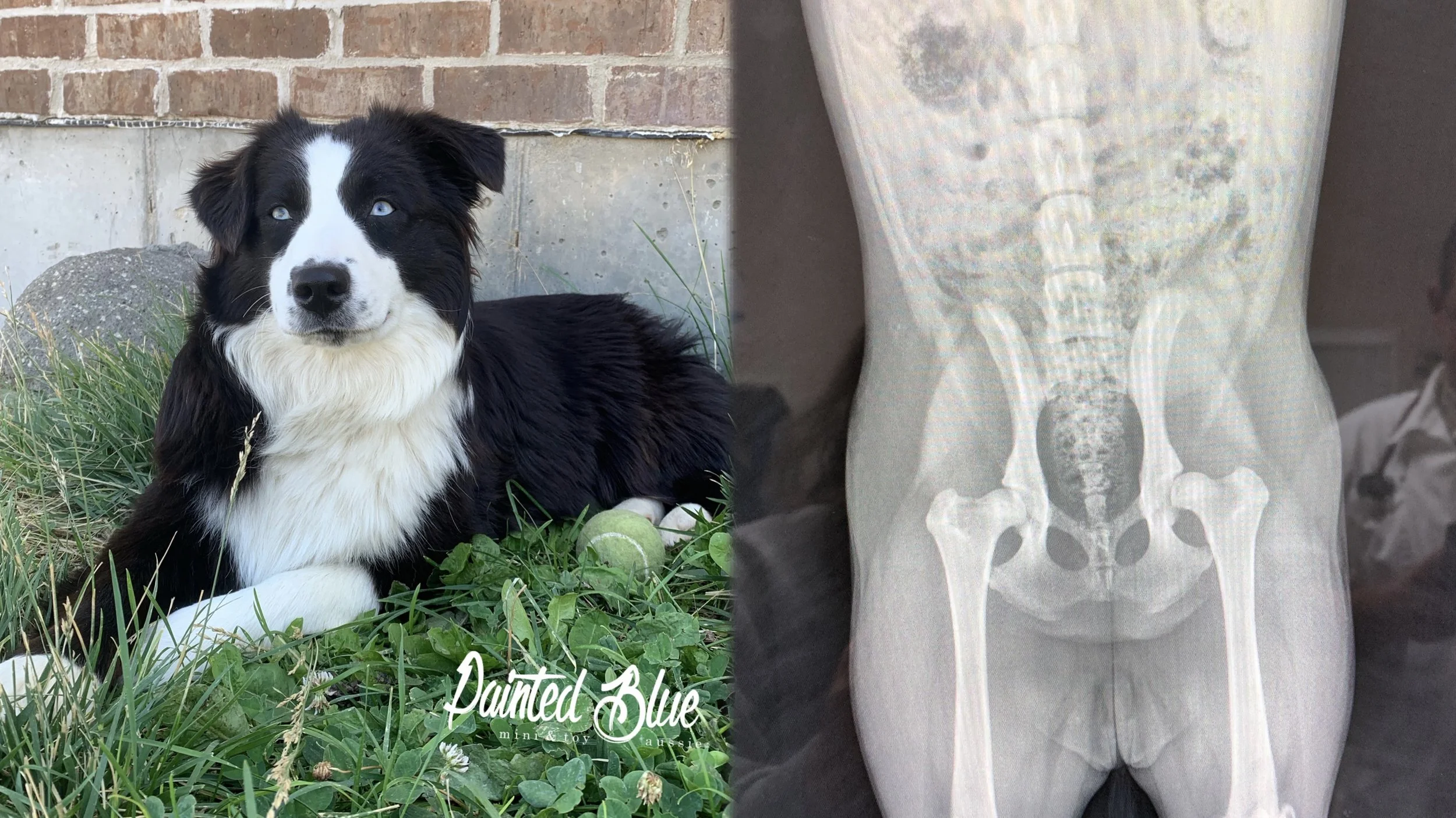
Crystal - Diagnosed with moderate to severe hip dysplasia at 6 months old. Crystal showed symptoms early, including difficulty standing after sitting, a bunny-hopping gait, and crying in pain when picked up awkwardly or jostled by other dogs.
The arthritic changes visible on an X-ray are often the end result of a process that began much earlier inside the joint.
5 - Looking Beneath the Surface: OFA & PennHIP
Because hip health cannot reliably be evaluated from the outside, breeders use radiographs to look more closely at the structure and stability of the hip joint. In the United States, OFA and PennHIP are two commonly used methods of hip screening.
Both use X-rays to evaluate the hips, but they do not evaluate them in exactly the same way or provide the same type of information. Understanding those differences is important when interpreting results and making breeding decisions.
OFA Hip Evaluation
The Orthopedic Foundation for Animals (OFA) evaluates hip conformation using a ventrodorsal, hip-extended radiograph. The dog is positioned on its back with the hind legs extended and rotated inward so the pelvis and both hip joints can be evaluated in a standardized position.
OFA does not base its grade on a single measurement. Instead, evaluators consider the overall appearance and relationship of the structures that make up the hip joint, looking for appropriate fit and congruity as well as signs of subluxation, remodeling or osteoarthritis.
Nine anatomical areas are specifically evaluated:
Craniolateral acetabular rim – the front and outer edge of the hip socket.
Cranial acetabular margin – the front margin of the socket.
Femoral head – the ball of the hip joint, including its shape and positioning.
Fovea capitis – the small normal depression on the femoral head where the ligament of the femoral head attaches.
Acetabular notch – the opening along the lower portion of the acetabulum.
Caudal acetabular rim – the rear margin of the hip socket.
Dorsal acetabular margin – the upper weight-bearing portion of the socket.
Junction of the femoral head and neck – evaluated for normal contour or evidence of thickening/remodeling.
Trochanteric fossa – the depression near the greater trochanter of the femur.
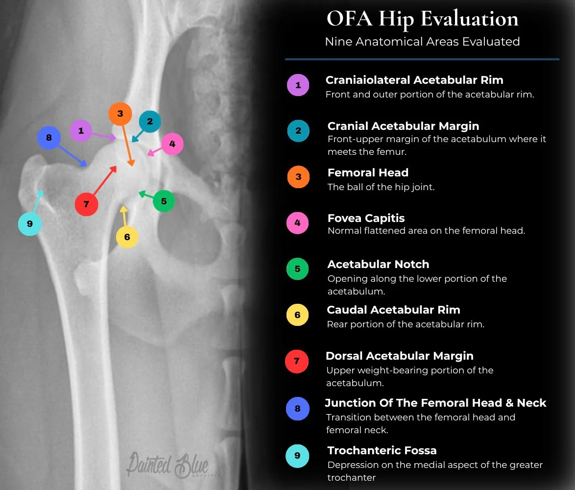
OFA then considers these findings together, particularly joint congruity, how well the femoral head is seated within the acetabulum, and whether there is evidence of degenerative joint disease, to assign the hip classification.
For dogs 24 months of age or older, the final classifications are Excellent, Good, Fair, Borderline, Mild, Moderate, or Severe. Excellent, Good and Fair are considered within the normal range for OFA certification; Mild, Moderate and Severe are dysplastic classifications. Dogs younger than 24 months can receive a preliminary evaluation rather than a final certification.
OFA is largely a subjective radiographic evaluation of phenotype and conformation performed by expert reviewers. PennHIP, by contrast, introduces an actual quantitative measurement of passive laxity, the Distraction Index.
Sources OFA Hip Screening & OFA The Use Of Health Databases And Selective Breeding
OFA doesn’t judge a hip by one feature alone. The final grade reflects the overall appearance, fit, and conformation of the joint across multiple anatomical areas.
PennHIP Evaluation
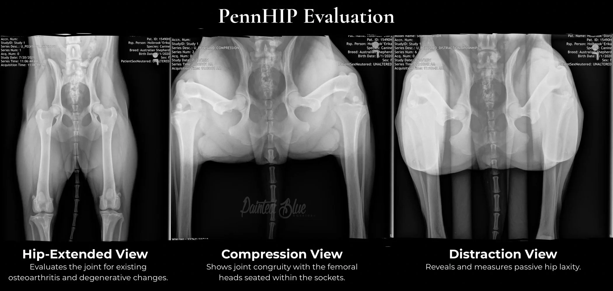
PennHIP takes a different approach to evaluating canine hips. Rather than relying on a single radiographic position, a PennHIP evaluation uses three separate X-ray views, with each providing different information about the joint.
Together, these views allow PennHIP to assess hip congruity, existing osteoarthritis, and passive hip laxity. A key feature of the method is its ability to objectively quantify laxity using the Distraction Index (DI), providing a numerical measurement of how much the femoral head can move within the acetabulum.
Hip-Extended View
In the hip-extended view, the dog is positioned on its back with the hind legs extended. This is similar to the traditional positioning used for an OFA radiograph. Within the PennHIP evaluation, this view is used primarily to look for existing radiographic signs of osteoarthritis or degenerative joint disease, such as remodeling or new bone formation around the joint.
Although the femoral heads may appear tightly seated in this position, extending the hips can tighten the joint capsule and mask some of the hip's underlying passive laxity. For that reason, PennHIP does not rely on the extended view alone to determine how loose the hips are.
Compression View
For the compression view, the hips are placed in a neutral position and the femoral heads are gently seated into the acetabula. This allows the evaluator to examine joint congruity, or how well the ball of the femoral head fits within the socket when it is fully seated.
This view also provides important anatomical landmarks used in PennHIP's measurements. A healthy, normally shaped joint should demonstrate a close, congruent fit when the femoral head is seated within the acetabulum. Remodeling associated with osteoarthritis can alter the shape of either surface and result in a poorer fit.
Distraction View
The distraction view is the feature that most clearly distinguishes PennHIP from conventional hip screening. The hips remain in a neutral position while a specially designed PennHIP distractor applies a controlled force that allows the femoral heads to move outward to the extent permitted by the joint's passive laxity.
This view reveals laxity that may not be visible on the hip-extended radiograph. PennHIP uses the displacement of the femoral head relative to the acetabulum to calculate the Distraction Index (DI), an objective numerical measurement of passive hip laxity. Lower DI values represent tighter hips, while higher values represent greater laxity.
Each PennHIP view answers a different question: Is there already osteoarthritis? How well does the joint fit when seated? And how much passive laxity is actually present?
Understanding the Distraction Index
The Distraction Index, or DI, is PennHIP's numerical measurement of passive hip laxity. It describes how far the center of the femoral head can move away from the center of the acetabulum when the hip is placed in the distraction position.
Each hip receives its own DI value. Lower numbers indicate tighter hips, while higher numbers indicate greater laxity. Unlike OFA grades, the DI exists on a continuous numerical scale rather than placing the hip into categories such as Excellent, Good, Fair, or dysplastic.
The DI is important because research has shown an association between greater passive hip laxity and an increased risk of developing hip osteoarthritis. However, the DI is a measure of laxity and risk, not a prediction of exactly what will happen to an individual dog. Dogs with similar DI values may not develop identical radiographic changes or clinical symptoms over their lifetimes. PennHIP: Measuring Hip Joint Laxity
Understanding A PennHIP Report
After the radiographs are submitted, PennHIP provides a Distraction Index (DI) for each hip along with an interpretation of the dog's results. The DI is a numerical measurement of passive hip laxity. Lower numbers represent tighter hips, while higher numbers represent greater laxity.
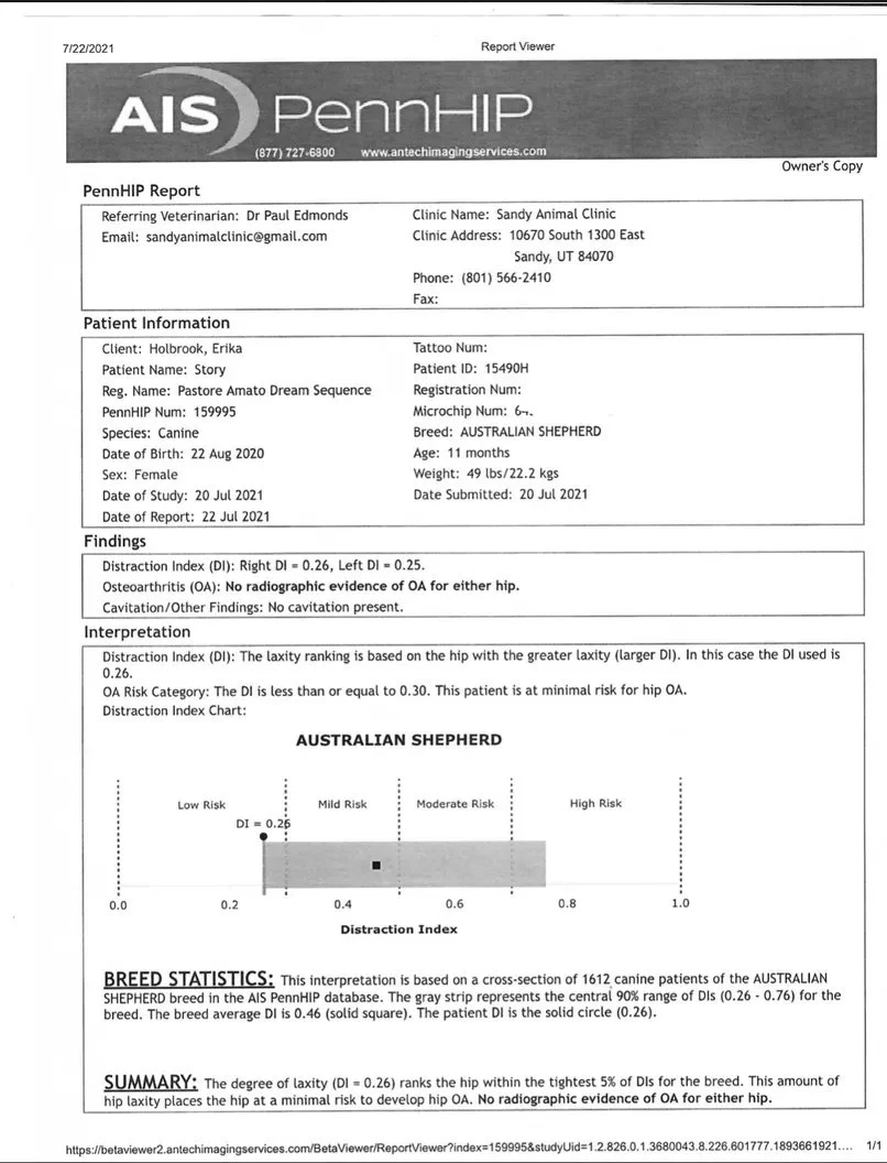
In the example of the dog (Story) x-rayed above, the dog's right hip received a DI of 0.26 and the left hip a DI of 0.25. PennHIP bases the laxity interpretation on the hip with the greater laxity, so the 0.26 DI is used for the overall interpretation.
The report placed this dog in the minimal risk category for developing hip osteoarthritis and noted that there was no radiographic evidence of osteoarthritis in either hip at the time of evaluation.
What Does The Chart Mean?
The chart provides additional context for interpreting the DI. In this report, the gray bar represents the central 90% range of Distraction Index values recorded for Australian Shepherds in the PennHIP database at that time. The square marks the breed's average DI, while the dog's result is shown by the solid circle.
This dog's DI of 0.26 placed her among the tightest 5% of Australian Shepherds in the database represented by this report. In other words, her hips demonstrated considerably less passive laxity than was typical for the breed population PennHIP had evaluated.
PennHIP is not simply a pass or fail test. The DI tells us how much passive laxity is present in each hip, while the rest of the report provides context about osteoarthritis risk and how that result compares with other dogs of the same breed.
6 - What Does This Mean For Breeding?
Hip screening gives breeders valuable information, but a hip score should never be interpreted in isolation. Canine hip dysplasia is a complex, multifactorial condition influenced by genetics as well as environmental factors, and no single radiograph can tell us everything about a dog's genetic contribution to the next generation.
We use OFA and PennHIP results as pieces of a much larger picture. We consider the individual dog's results alongside pedigree, relatives, previous offspring when available, overall health, structure, temperament, genetic diversity, and the strengths and weaknesses of the proposed pairing.
A less-than-ideal result does not automatically mean that a dog has nothing valuable to contribute to a breeding program, just as an Excellent OFA rating or very low PennHIP DI does not guarantee that every puppy produced will have healthy hips. The purpose of testing is to give us information we can use to make thoughtful, informed breeding decisions and to continually work toward improving the next generation.
For us, responsible breeding is not about chasing a single number or result. It is about understanding the information we have, preserving what is valuable, and making each pairing with intention.
Health testing does not eliminate every risk. It gives us better information with which to make better decisions.